Textile Documents › Authors › Author: E. R. Schwarz
3 documents found
Sort: 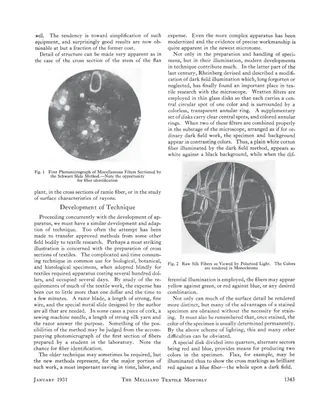
Newer Developments in Textile Microscopy
Article · 5 pages
A 1931 paper by E. R. Schwarz of M. I. T. surveying new developments in textile microscopy, presented to ASTM Committee D-13 in October 1930. It covers specialized microscopes and mounting and measuring equipment, a quick low-cost method for cutting fiber cross sections (the Schwarz slide), Rheinberg colored dark-field illumination, and polarized light. It also discusses the handling and statistical treatment of data, applied mill research with pocket and stereoscopic microscopes, and prospects for x-ray, fluorescence, and photoelectric methods. Illustrated with seven photomicrographs and apparatus figures.
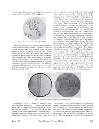
Wool, Woolens, and Worsted Through the Microscope, Part 1
Article · 3 pages
Part 1 of an article by Edward R. Schwarz on using the microscope to study wool fibers and wool fabrics. It argues that attempts at artificial wool fail to account for the fiber's complex structure, and that manufacturers, like wool growers, should test fibers. It describes the cuticle, scales, cortex and medulla, then reviews research on fineness, spinning quality, crimp, grading, felting and chlorination. Two photomicrograph figures compare imitation and real wool fibers, and a numbered reference list closes the piece.
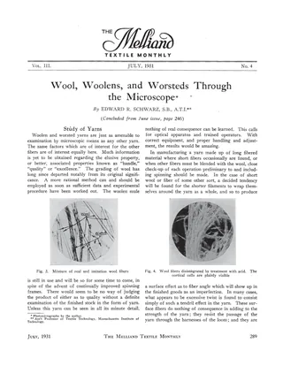
Wool, Woolens, and Worsted Through the Microscope, Part 2
Article · 4 pages
Concluding part of an article by Edward R. Schwarz of MIT on using the microscope to examine wool, woolen and worsted yarns and fabrics. It covers yarn study (surface fibers, twist faults, rayon blends), checking double cloths and backed fabrics against their designs, and using the stereoscopic microscope on pile fabrics such as Wilton carpet and mohair plush. It closes with remarks on mill quality inspection and the need for research-trained laboratory staff. Illustrated with photomicrographs (Figs. 3-11).